fluorescent semiconductor nanoparticle quantum dot Search Results


90
Alomone Labs qd ngf
( a ) Partial sequence of the 3′-UTR of rat Pafah1b1 starting at the stop codon (*). The binding regions of the CUGU and control LNAs are indicated in maroon and grey, respectively. The CUGU element is underlined. ( b ) Dissociated DRG were transfected with control and CUGU LNA, and 24 h later, APC RNA immunoprecipitation was performed. Pafah1b1 was quantified by RT–PCR. 2 −ΔΔCT values are reported relative to Tubb3 (positive control, binds APC but is not targeted by the LNAs). Gfp was included as a control (no reads detected). Means±s.e.m. ( n =2 biological replicates with two technical replicates each). * P ≥0.05. t -test. ( c ) DRG neurons were cultured in microfluidic chambers. On DIV 3, the <t>NGF</t> concentration in the axonal chamber was changed to 5 ng ml −1 , and cell bodies were selectively transfected with the control or CUGU LNAs. Twenty-four hours after transfection, axons were treated with 0, 5 or 100 ng ml −1 NGF for 12 h, and axonal Pafah1b1 mRNA levels were determined by FISH. Background fluorescence was determined using a Gfp probe and subtracted. Means±s.e.m. of 15 optical fields per condition ( n =3 biological replicates). * P ≥0.05. Two-way ANOVA with Fisher's least significant difference test. Scale bar, 5 μm. ( d ) DRG neurons were cultured and transfected as in a . Twenty-four hours after transfection, axons were treated with 0, 5 or 100 ng ml −1 NGF for 10 min, and axonal Lis1 protein levels were measured by quantitative immunofluorescence. Means±s.e.m. of 20–30 optical fields per conditions ( n =4–6 biological replicates). * P ≥0.05. Two-way ANOVA with Fisher's LSD test. ( e – g ) DRG neurons were cultured and transfected as in a . Twenty-four hours after <t>transfection,</t> <t>transport</t> of LysoTracker-positive particles was observed in axons at baseline NGF ( e ), without NGF ( f ) or stimulated with NGF ( g ). Live-imaging time-lapse series of axonal fields were acquired, with images being taken every 13 s for 4 min. LysoTracker-positive particles with diameters ≥1 μm were scored as anterograde, retrograde, bidirectional or stationary. Means±s.e.m. of nine optical fields per conditions ( n =3 biological replicates). ** P ≥0.01. One-way ANOVA with Bonferroni's multiple comparisons test. NS, not significant.
Qd Ngf, supplied by Alomone Labs, used in various techniques. Bioz Stars score: 90/100, based on 1 PubMed citations. ZERO BIAS - scores, article reviews, protocol conditions and more
https://www.bioz.com/result/qd ngf/product/Alomone Labs
Average 90 stars, based on 1 article reviews
qd ngf - by Bioz Stars, 2026-05
90/100 stars
  Buy from Supplier

99
Thermo Fisher collagen 1α promoter
( a ) Partial sequence of the 3′-UTR of rat Pafah1b1 starting at the stop codon (*). The binding regions of the CUGU and control LNAs are indicated in maroon and grey, respectively. The CUGU element is underlined. ( b ) Dissociated DRG were transfected with control and CUGU LNA, and 24 h later, APC RNA immunoprecipitation was performed. Pafah1b1 was quantified by RT–PCR. 2 −ΔΔCT values are reported relative to Tubb3 (positive control, binds APC but is not targeted by the LNAs). Gfp was included as a control (no reads detected). Means±s.e.m. ( n =2 biological replicates with two technical replicates each). * P ≥0.05. t -test. ( c ) DRG neurons were cultured in microfluidic chambers. On DIV 3, the <t>NGF</t> concentration in the axonal chamber was changed to 5 ng ml −1 , and cell bodies were selectively transfected with the control or CUGU LNAs. Twenty-four hours after transfection, axons were treated with 0, 5 or 100 ng ml −1 NGF for 12 h, and axonal Pafah1b1 mRNA levels were determined by FISH. Background fluorescence was determined using a Gfp probe and subtracted. Means±s.e.m. of 15 optical fields per condition ( n =3 biological replicates). * P ≥0.05. Two-way ANOVA with Fisher's least significant difference test. Scale bar, 5 μm. ( d ) DRG neurons were cultured and transfected as in a . Twenty-four hours after transfection, axons were treated with 0, 5 or 100 ng ml −1 NGF for 10 min, and axonal Lis1 protein levels were measured by quantitative immunofluorescence. Means±s.e.m. of 20–30 optical fields per conditions ( n =4–6 biological replicates). * P ≥0.05. Two-way ANOVA with Fisher's LSD test. ( e – g ) DRG neurons were cultured and transfected as in a . Twenty-four hours after <t>transfection,</t> <t>transport</t> of LysoTracker-positive particles was observed in axons at baseline NGF ( e ), without NGF ( f ) or stimulated with NGF ( g ). Live-imaging time-lapse series of axonal fields were acquired, with images being taken every 13 s for 4 min. LysoTracker-positive particles with diameters ≥1 μm were scored as anterograde, retrograde, bidirectional or stationary. Means±s.e.m. of nine optical fields per conditions ( n =3 biological replicates). ** P ≥0.01. One-way ANOVA with Bonferroni's multiple comparisons test. NS, not significant.
Collagen 1α Promoter, supplied by Thermo Fisher, used in various techniques. Bioz Stars score: 99/100, based on 1 PubMed citations. ZERO BIAS - scores, article reviews, protocol conditions and more
https://www.bioz.com/result/collagen 1α promoter/product/Thermo Fisher
Average 99 stars, based on 1 article reviews
collagen 1α promoter - by Bioz Stars, 2026-05
99/100 stars
  Buy from Supplier

90
Reagen LLC graphene quantum dots
( a ) Partial sequence of the 3′-UTR of rat Pafah1b1 starting at the stop codon (*). The binding regions of the CUGU and control LNAs are indicated in maroon and grey, respectively. The CUGU element is underlined. ( b ) Dissociated DRG were transfected with control and CUGU LNA, and 24 h later, APC RNA immunoprecipitation was performed. Pafah1b1 was quantified by RT–PCR. 2 −ΔΔCT values are reported relative to Tubb3 (positive control, binds APC but is not targeted by the LNAs). Gfp was included as a control (no reads detected). Means±s.e.m. ( n =2 biological replicates with two technical replicates each). * P ≥0.05. t -test. ( c ) DRG neurons were cultured in microfluidic chambers. On DIV 3, the <t>NGF</t> concentration in the axonal chamber was changed to 5 ng ml −1 , and cell bodies were selectively transfected with the control or CUGU LNAs. Twenty-four hours after transfection, axons were treated with 0, 5 or 100 ng ml −1 NGF for 12 h, and axonal Pafah1b1 mRNA levels were determined by FISH. Background fluorescence was determined using a Gfp probe and subtracted. Means±s.e.m. of 15 optical fields per condition ( n =3 biological replicates). * P ≥0.05. Two-way ANOVA with Fisher's least significant difference test. Scale bar, 5 μm. ( d ) DRG neurons were cultured and transfected as in a . Twenty-four hours after transfection, axons were treated with 0, 5 or 100 ng ml −1 NGF for 10 min, and axonal Lis1 protein levels were measured by quantitative immunofluorescence. Means±s.e.m. of 20–30 optical fields per conditions ( n =4–6 biological replicates). * P ≥0.05. Two-way ANOVA with Fisher's LSD test. ( e – g ) DRG neurons were cultured and transfected as in a . Twenty-four hours after <t>transfection,</t> <t>transport</t> of LysoTracker-positive particles was observed in axons at baseline NGF ( e ), without NGF ( f ) or stimulated with NGF ( g ). Live-imaging time-lapse series of axonal fields were acquired, with images being taken every 13 s for 4 min. LysoTracker-positive particles with diameters ≥1 μm were scored as anterograde, retrograde, bidirectional or stationary. Means±s.e.m. of nine optical fields per conditions ( n =3 biological replicates). ** P ≥0.01. One-way ANOVA with Bonferroni's multiple comparisons test. NS, not significant.
Graphene Quantum Dots, supplied by Reagen LLC, used in various techniques. Bioz Stars score: 90/100, based on 1 PubMed citations. ZERO BIAS - scores, article reviews, protocol conditions and more
https://www.bioz.com/result/graphene quantum dots/product/Reagen LLC
Average 90 stars, based on 1 article reviews
graphene quantum dots - by Bioz Stars, 2026-05
90/100 stars
  Buy from Supplier

99
Thermo Fisher streptavidin conjugated quantum dots
(A) Schematic diagram depicting a <t>streptavidin-conjugated</t> quantum dot (QD) conjugated to biotinylated antibodies against the luminal domain of VGAT. (B) Colocalization of VGAT-QD‒loaded inhibitory vesicles (green) and CypHer5E-VGAT‒labeled presynaptic boutons (red) in cultured hippocampal neurons. Scale bar: 1 µm. (C) Three-dimensional trajectory of a VGAT-QD‒loaded inhibitory vesicle overlaid on the x - y plane of a CypHer5E-VGAT‒ labeled presynaptic bouton. The color bar represents elapsed time; electrical stimulation (10 Hz) started at 20 s, and the vesicle underwent exocytosis at 32.0 s. (D) Fluorescence images of the VGAT-QD‒loaded vesicle shown in panel C taken at the indicated times. Scale bar: 0.5 µm. (E) Three-dimensional position, radial distance from the momentary position to the fusion site (R), and fluorescence intensity (F) of the VGAT-QD‒loaded vesicle shown in panel C. Note the photoblinking events (e.g., at approximately 8 s, 13 s and 15 s), confirming the presence of one QD inside the vesicle. Electrical stimuli (10 Hz) were applied for 120 s starting at 20 s (green horizontal bar).
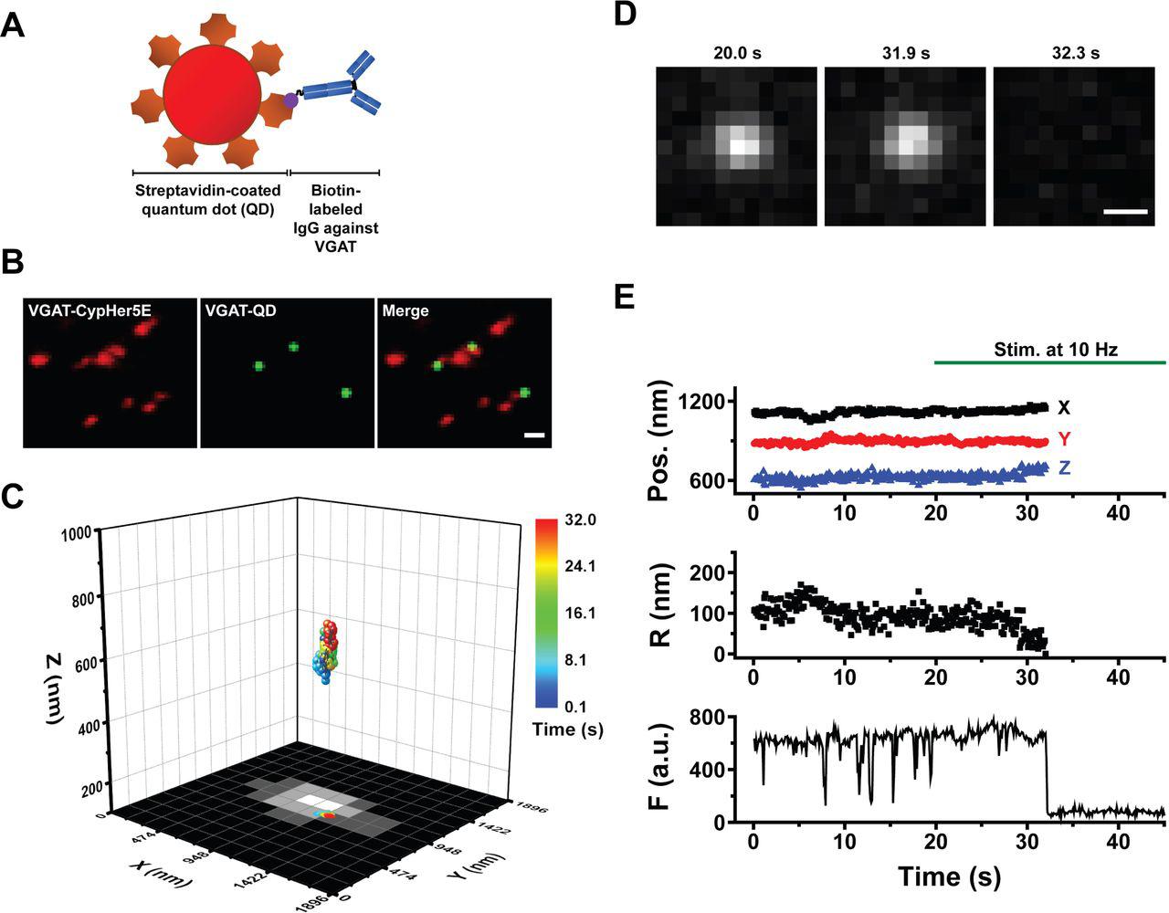
Streptavidin Conjugated Quantum Dots, supplied by Thermo Fisher, used in various techniques. Bioz Stars score: 99/100, based on 1 PubMed citations. ZERO BIAS - scores, article reviews, protocol conditions and more
https://www.bioz.com/result/streptavidin conjugated quantum dots/product/Thermo Fisher
Average 99 stars, based on 1 article reviews
streptavidin conjugated quantum dots - by Bioz Stars, 2026-05
99/100 stars
  Buy from Supplier

99
Thermo Fisher facs buffer

Facs Buffer, supplied by Thermo Fisher, used in various techniques. Bioz Stars score: 99/100, based on 1 PubMed citations. ZERO BIAS - scores, article reviews, protocol conditions and more
https://www.bioz.com/result/facs buffer/product/Thermo Fisher
Average 99 stars, based on 1 article reviews
facs buffer - by Bioz Stars, 2026-05
99/100 stars
  Buy from Supplier

90
Ocean NanoTech green fluorescent quantum dots (qds

Green Fluorescent Quantum Dots (Qds, supplied by Ocean NanoTech, used in various techniques. Bioz Stars score: 90/100, based on 1 PubMed citations. ZERO BIAS - scores, article reviews, protocol conditions and more
https://www.bioz.com/result/green fluorescent quantum dots (qds/product/Ocean NanoTech
Average 90 stars, based on 1 article reviews
green fluorescent quantum dots (qds - by Bioz Stars, 2026-05
90/100 stars
  Buy from Supplier

90
BioApplications Inc fluorescent semiconductor nanocrystals

Fluorescent Semiconductor Nanocrystals, supplied by BioApplications Inc, used in various techniques. Bioz Stars score: 90/100, based on 1 PubMed citations. ZERO BIAS - scores, article reviews, protocol conditions and more
https://www.bioz.com/result/fluorescent semiconductor nanocrystals/product/BioApplications Inc
Average 90 stars, based on 1 article reviews
fluorescent semiconductor nanocrystals - by Bioz Stars, 2026-05
90/100 stars
  Buy from Supplier

90
AnaSpec fluorescent probes and quantum dots

Fluorescent Probes And Quantum Dots, supplied by AnaSpec, used in various techniques. Bioz Stars score: 90/100, based on 1 PubMed citations. ZERO BIAS - scores, article reviews, protocol conditions and more
https://www.bioz.com/result/fluorescent probes and quantum dots/product/AnaSpec
Average 90 stars, based on 1 article reviews
fluorescent probes and quantum dots - by Bioz Stars, 2026-05
90/100 stars
  Buy from Supplier

90
Keerthana Industries pyrene carboxaldehyde-carbon quantum dots (pc-cd) fluorescent nanosensor

Pyrene Carboxaldehyde Carbon Quantum Dots (Pc Cd) Fluorescent Nanosensor, supplied by Keerthana Industries, used in various techniques. Bioz Stars score: 90/100, based on 1 PubMed citations. ZERO BIAS - scores, article reviews, protocol conditions and more
https://www.bioz.com/result/pyrene carboxaldehyde-carbon quantum dots (pc-cd) fluorescent nanosensor/product/Keerthana Industries
Average 90 stars, based on 1 article reviews
pyrene carboxaldehyde-carbon quantum dots (pc-cd) fluorescent nanosensor - by Bioz Stars, 2026-05
90/100 stars
  Buy from Supplier

90
Optik GmbH fluorescent graphene quantum dots

Fluorescent Graphene Quantum Dots, supplied by Optik GmbH, used in various techniques. Bioz Stars score: 90/100, based on 1 PubMed citations. ZERO BIAS - scores, article reviews, protocol conditions and more
https://www.bioz.com/result/fluorescent graphene quantum dots/product/Optik GmbH
Average 90 stars, based on 1 article reviews
fluorescent graphene quantum dots - by Bioz Stars, 2026-05
90/100 stars
  Buy from Supplier

99
BMG Labtech fluostar omega fluorescence microplate reader

Fluostar Omega Fluorescence Microplate Reader, supplied by BMG Labtech, used in various techniques. Bioz Stars score: 99/100, based on 1 PubMed citations. ZERO BIAS - scores, article reviews, protocol conditions and more
https://www.bioz.com/result/fluostar omega fluorescence microplate reader/product/BMG Labtech
Average 99 stars, based on 1 article reviews
fluostar omega fluorescence microplate reader - by Bioz Stars, 2026-05
99/100 stars
  Buy from Supplier

90
Becton Dickinson cd4 (ox 35

Cd4 (Ox 35, supplied by Becton Dickinson, used in various techniques. Bioz Stars score: 90/100, based on 1 PubMed citations. ZERO BIAS - scores, article reviews, protocol conditions and more
https://www.bioz.com/result/cd4 (ox 35/product/Becton Dickinson
Average 90 stars, based on 1 article reviews
cd4 (ox 35 - by Bioz Stars, 2026-05
90/100 stars
  Buy from Supplier

Image Search Results


( a ) Partial sequence of the 3′-UTR of rat Pafah1b1 starting at the stop codon (*). The binding regions of the CUGU and control LNAs are indicated in maroon and grey, respectively. The CUGU element is underlined. ( b ) Dissociated DRG were transfected with control and CUGU LNA, and 24 h later, APC RNA immunoprecipitation was performed. Pafah1b1 was quantified by RT–PCR. 2 −ΔΔCT values are reported relative to Tubb3 (positive control, binds APC but is not targeted by the LNAs). Gfp was included as a control (no reads detected). Means±s.e.m. ( n =2 biological replicates with two technical replicates each). * P ≥0.05. t -test. ( c ) DRG neurons were cultured in microfluidic chambers. On DIV 3, the NGF concentration in the axonal chamber was changed to 5 ng ml −1 , and cell bodies were selectively transfected with the control or CUGU LNAs. Twenty-four hours after transfection, axons were treated with 0, 5 or 100 ng ml −1 NGF for 12 h, and axonal Pafah1b1 mRNA levels were determined by FISH. Background fluorescence was determined using a Gfp probe and subtracted. Means±s.e.m. of 15 optical fields per condition ( n =3 biological replicates). * P ≥0.05. Two-way ANOVA with Fisher's least significant difference test. Scale bar, 5 μm. ( d ) DRG neurons were cultured and transfected as in a . Twenty-four hours after transfection, axons were treated with 0, 5 or 100 ng ml −1 NGF for 10 min, and axonal Lis1 protein levels were measured by quantitative immunofluorescence. Means±s.e.m. of 20–30 optical fields per conditions ( n =4–6 biological replicates). * P ≥0.05. Two-way ANOVA with Fisher's LSD test. ( e – g ) DRG neurons were cultured and transfected as in a . Twenty-four hours after transfection, transport of LysoTracker-positive particles was observed in axons at baseline NGF ( e ), without NGF ( f ) or stimulated with NGF ( g ). Live-imaging time-lapse series of axonal fields were acquired, with images being taken every 13 s for 4 min. LysoTracker-positive particles with diameters ≥1 μm were scored as anterograde, retrograde, bidirectional or stationary. Means±s.e.m. of nine optical fields per conditions ( n =3 biological replicates). ** P ≥0.01. One-way ANOVA with Bonferroni's multiple comparisons test. NS, not significant.

Journal: Nature Communications

Article Title: Local synthesis of dynein cofactors matches retrograde transport to acutely changing demands

doi: 10.1038/ncomms13865

Figure Lengend Snippet: ( a ) Partial sequence of the 3′-UTR of rat Pafah1b1 starting at the stop codon (*). The binding regions of the CUGU and control LNAs are indicated in maroon and grey, respectively. The CUGU element is underlined. ( b ) Dissociated DRG were transfected with control and CUGU LNA, and 24 h later, APC RNA immunoprecipitation was performed. Pafah1b1 was quantified by RT–PCR. 2 −ΔΔCT values are reported relative to Tubb3 (positive control, binds APC but is not targeted by the LNAs). Gfp was included as a control (no reads detected). Means±s.e.m. ( n =2 biological replicates with two technical replicates each). * P ≥0.05. t -test. ( c ) DRG neurons were cultured in microfluidic chambers. On DIV 3, the NGF concentration in the axonal chamber was changed to 5 ng ml −1 , and cell bodies were selectively transfected with the control or CUGU LNAs. Twenty-four hours after transfection, axons were treated with 0, 5 or 100 ng ml −1 NGF for 12 h, and axonal Pafah1b1 mRNA levels were determined by FISH. Background fluorescence was determined using a Gfp probe and subtracted. Means±s.e.m. of 15 optical fields per condition ( n =3 biological replicates). * P ≥0.05. Two-way ANOVA with Fisher's least significant difference test. Scale bar, 5 μm. ( d ) DRG neurons were cultured and transfected as in a . Twenty-four hours after transfection, axons were treated with 0, 5 or 100 ng ml −1 NGF for 10 min, and axonal Lis1 protein levels were measured by quantitative immunofluorescence. Means±s.e.m. of 20–30 optical fields per conditions ( n =4–6 biological replicates). * P ≥0.05. Two-way ANOVA with Fisher's LSD test. ( e – g ) DRG neurons were cultured and transfected as in a . Twenty-four hours after transfection, transport of LysoTracker-positive particles was observed in axons at baseline NGF ( e ), without NGF ( f ) or stimulated with NGF ( g ). Live-imaging time-lapse series of axonal fields were acquired, with images being taken every 13 s for 4 min. LysoTracker-positive particles with diameters ≥1 μm were scored as anterograde, retrograde, bidirectional or stationary. Means±s.e.m. of nine optical fields per conditions ( n =3 biological replicates). ** P ≥0.01. One-way ANOVA with Bonferroni's multiple comparisons test. NS, not significant.

Article Snippet: For imaging transport of NGF-containing endosomes, QD-NGF was prepared by mixing mouse NGF 2.5S-Biotin (Alomone Labs, Jerusalem) and Qdot 585 Streptavidin Conjugate in a 1:1.2 molar ratio, and incubating them together at 4 °C with continuous inversion for 24 h. QD-NGF was diluted to 100 ng ml −1 and added to axons with a medium change 15 min before imaging.

Techniques: Sequencing, Binding Assay, Transfection, Immunoprecipitation, Reverse Transcription Polymerase Chain Reaction, Positive Control, Cell Culture, Concentration Assay, Fluorescence, Immunofluorescence, Imaging

(A) Schematic diagram depicting a streptavidin-conjugated quantum dot (QD) conjugated to biotinylated antibodies against the luminal domain of VGAT. (B) Colocalization of VGAT-QD‒loaded inhibitory vesicles (green) and CypHer5E-VGAT‒labeled presynaptic boutons (red) in cultured hippocampal neurons. Scale bar: 1 µm. (C) Three-dimensional trajectory of a VGAT-QD‒loaded inhibitory vesicle overlaid on the x - y plane of a CypHer5E-VGAT‒ labeled presynaptic bouton. The color bar represents elapsed time; electrical stimulation (10 Hz) started at 20 s, and the vesicle underwent exocytosis at 32.0 s. (D) Fluorescence images of the VGAT-QD‒loaded vesicle shown in panel C taken at the indicated times. Scale bar: 0.5 µm. (E) Three-dimensional position, radial distance from the momentary position to the fusion site (R), and fluorescence intensity (F) of the VGAT-QD‒loaded vesicle shown in panel C. Note the photoblinking events (e.g., at approximately 8 s, 13 s and 15 s), confirming the presence of one QD inside the vesicle. Electrical stimuli (10 Hz) were applied for 120 s starting at 20 s (green horizontal bar).

Journal: bioRxiv

Article Title: Inhibitory synaptic vesicles have unique dynamics and exocytosis properties

doi: 10.1101/2020.09.21.289314

Figure Lengend Snippet: (A) Schematic diagram depicting a streptavidin-conjugated quantum dot (QD) conjugated to biotinylated antibodies against the luminal domain of VGAT. (B) Colocalization of VGAT-QD‒loaded inhibitory vesicles (green) and CypHer5E-VGAT‒labeled presynaptic boutons (red) in cultured hippocampal neurons. Scale bar: 1 µm. (C) Three-dimensional trajectory of a VGAT-QD‒loaded inhibitory vesicle overlaid on the x - y plane of a CypHer5E-VGAT‒ labeled presynaptic bouton. The color bar represents elapsed time; electrical stimulation (10 Hz) started at 20 s, and the vesicle underwent exocytosis at 32.0 s. (D) Fluorescence images of the VGAT-QD‒loaded vesicle shown in panel C taken at the indicated times. Scale bar: 0.5 µm. (E) Three-dimensional position, radial distance from the momentary position to the fusion site (R), and fluorescence intensity (F) of the VGAT-QD‒loaded vesicle shown in panel C. Note the photoblinking events (e.g., at approximately 8 s, 13 s and 15 s), confirming the presence of one QD inside the vesicle. Electrical stimuli (10 Hz) were applied for 120 s starting at 20 s (green horizontal bar).

Article Snippet: The biotinylated monoclonal mouse anti-Syt1 antibody (105 311BT, Synaptic Systems) or the biotinylated anti-VGAT antibody (131 103CpH, Synaptic Systems) was conjugated to streptavidin-conjugated quantum dots (cat. A10196, Thermo Fisher Scientific), and vesicles were loaded as described previously ( ).

Techniques: Cell Culture, Labeling, Fluorescence

Journal: iScience

Article Title: Preoperative immune checkpoint inhibition and cryoablation in early-stage breast cancer

doi: 10.1016/j.isci.2024.108880

Figure Lengend Snippet:

Article Snippet: Briefly, one million PBMCs and TILs were washed with 2 mL FACS buffer (PBS [phosphate-buffered saline] containing bovine 1% serum albumin and 0.05 mM EDTA), resuspended in 50 μL FACS buffer and stained with a fixable Aqua viability dye (Invitrogen) and a cocktail of antibodies to the following surface markers: CD8-Qdot 605 (Invitrogen, 3B5), CD4-Qdot 655 (Invitrogen, S3.5), PD-1-PE (BD, MIH4), LAG-3-FITC (Enzo Life Sciences, 17B4), ICOS-PE-Cy7 (eBioscience, ISA-3), TIM-3-APC (R&D Systems, 344823).

Techniques: Recombinant, Saline, Staining, Extraction, Software, Flow Cytometry